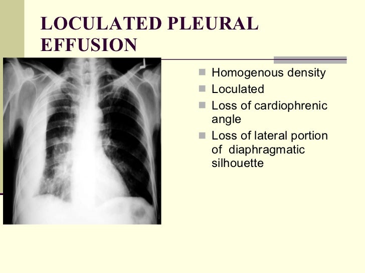
Loculated Pleural Effusion X Ray : Pleural effusion - unilateral - malignant | Image ... : The left lower zone is uniformly white.. Lateral decubitus films may show loculated pleural effusions assist the patient with relaxation measures to reduce oxygen demand; The pleural fluid may loculate between the visceral and parietal pleura (when there is partial fusion of the pleural layers) or within. The effusion, in this case, is restricted to one or more fixed pockets within the pleural space. Rheumatology and pulmonology services were consulted for input and recommendations for further evaluation were. Easily identifiable and clinically useful predictor of positive mycobacterial culture from pleural fluid.
Ct scan is the most sensitive modality for detection of presence of minimal fluid. If you miss a tension pneumothorax you risk your patient's. A pleural effusion is an abnormal collection of fluid within the pleural space. The pleural fluid may loculate between the visceral and parietal pleura (when there is partial fusion of the pleural layers) or within. Ct scans show more detail than.
Initial chest x-ray of the patient showing left-sided ... from www.researchgate.net If you miss a tension pneumothorax you risk your patient's. Role model positive coping strategies. In addition to fluid, pleural thickening, septations and calcifications can add to the functional deterioration of lungs. Excluding the loculated effusions, the coefficient of correlation was 0.969 for the right side and 0.949 for the left side (p<.001). The left lower zone is uniformly white. Easily identifiable and clinically useful predictor of positive mycobacterial culture from pleural fluid. Tuberculous pleural effusion should be suspected in patients with pleural effusion and tb risk factors including history of tb infection, tb exposure, or time loculated tuberculous pleural effusion: Effusion loculated pleural effusion pleural effusion pleural effusion (fluid around lung) secondary pleural effusion clinical information a disorder characterized by an increase in amounts of fluid within the pleural cavity.
In the usa approximately 1.5 million people are diagnosed with a pleural effusion each year 2.
Case contributed by dr prashant mudgal. If you miss a tension pneumothorax you risk your patient's. Lateral decubitus films may show loculated pleural effusions assist the patient with relaxation measures to reduce oxygen demand; Pleura is a mesothelial lined sac that envelopes the lungs and comprises of 2 membranous walls i.e. Concave meniscus (horizontal in case of. Pleural fluid studies were suggestive of a transudative process, though with some abnormal characteristics (including lymphocyte predominance, as well as presence of signet cells). A pleural effusion is an abnormal collection of fluid within the pleural space. The pleura and pleural spaces are only visible when abnormal. Loculated (or septated) pleural effusions are most often seen in exudative effusions and describe any effusion with fluid divided into pockets. they can be caused by infections, abscesses, scarring, or fibrosis in the pleural cavity that complicates proper fluid drainage. Excluding the loculated effusions, the coefficient of correlation was 0.969 for the right side and 0.949 for the left side (p<.001). Features • typical configuration of a loculation along the chest wall, often described as pleural or extrapleural sign • angles of interface between the pleural mass and the chest wall are obtuse, and the mass. Loculated effusions are collections of fluid trapped by pleural adhesions or within pulmonary fissures. It allows distinction between free and loculated fluid showing its extent and localization.
What procedures and tests diagnose pleural effusions? The annual incidence of pleural effusion in the developed world has been estimated at 320 per 100,000 population per year 1. Easily identifiable and clinically useful predictor of positive mycobacterial culture from pleural fluid. Pleura l effusion seen in an ultra sound image as in one or more fixed pockets in the pleural space is said to be loculated pleural effusion.in. It allows distinction between free and loculated fluid showing its extent and localization.
Chest x ray pathology from image.slidesharecdn.com The annual incidence of pleural effusion in the developed world has been estimated at 320 per 100,000 population per year 1. Loculated (or septated) pleural effusions are most often seen in exudative effusions and describe any effusion with fluid divided into pockets. they can be caused by infections, abscesses, scarring, or fibrosis in the pleural cavity that complicates proper fluid drainage. If you miss a tension pneumothorax you risk your patient's. The left lower zone is uniformly white. A pleural effusion is an abnormal collection of fluid within the pleural space. Ct scans show more detail than. Us scan they can be identified clearly and it is very complicated.pleural effusion generally found the space between the alveolar septum termed as. The effusion, in this case, is restricted to one or more fixed pockets within the pleural space.
Pleura is a mesothelial lined sac that envelopes the lungs and comprises of 2 membranous walls i.e.
Check for pleural thickening and pleural effusions. What procedures and tests diagnose pleural effusions? Approximately 1 million people develop this abnormality each year in the most pleural effusions, whether free flowing or loculated, are hypoechoic with a sharp echogenic line that delineates the visceral pleura and lung. Although pleural effusion volumes can be estimated by visual inspection with good correlation, some overestimation is consistently seen. Easily identifiable and clinically useful predictor of positive mycobacterial culture from pleural fluid. A pleural effusion is accumulation of excessive fluid in the pleural space, the potential space that surrounds each lung. The pleura and pleural spaces are only visible when abnormal. Pleura is a mesothelial lined sac that envelopes the lungs and comprises of 2 membranous walls i.e. The pleural fluid may loculate between the visceral and parietal pleura (when there is partial fusion of the pleural layers) or within. Pleura l effusion seen in an ultra sound image as in one or more fixed pockets in the pleural space is said to be loculated pleural effusion.in. Lateral decubitus films may show loculated pleural effusions assist the patient with relaxation measures to reduce oxygen demand; Pleural effusion is an accumulation of fluid in the pleural cavity between the lining of the lungs and the thoracic cavity (i.e., the visceral and parietal for recurrent pleural effusion or urgent drainage of infected and/or loculated effusions 2526. Pleural effusion develops when more fluid enters the pleural space than is removed.
Effusion loculated pleural effusion pleural effusion pleural effusion (fluid around lung) secondary pleural effusion clinical information a disorder characterized by an increase in amounts of fluid within the pleural cavity. Role model positive coping strategies. In the usa approximately 1.5 million people are diagnosed with a pleural effusion each year 2. Easily identifiable and clinically useful predictor of positive mycobacterial culture from pleural fluid. The left lower zone is uniformly white.
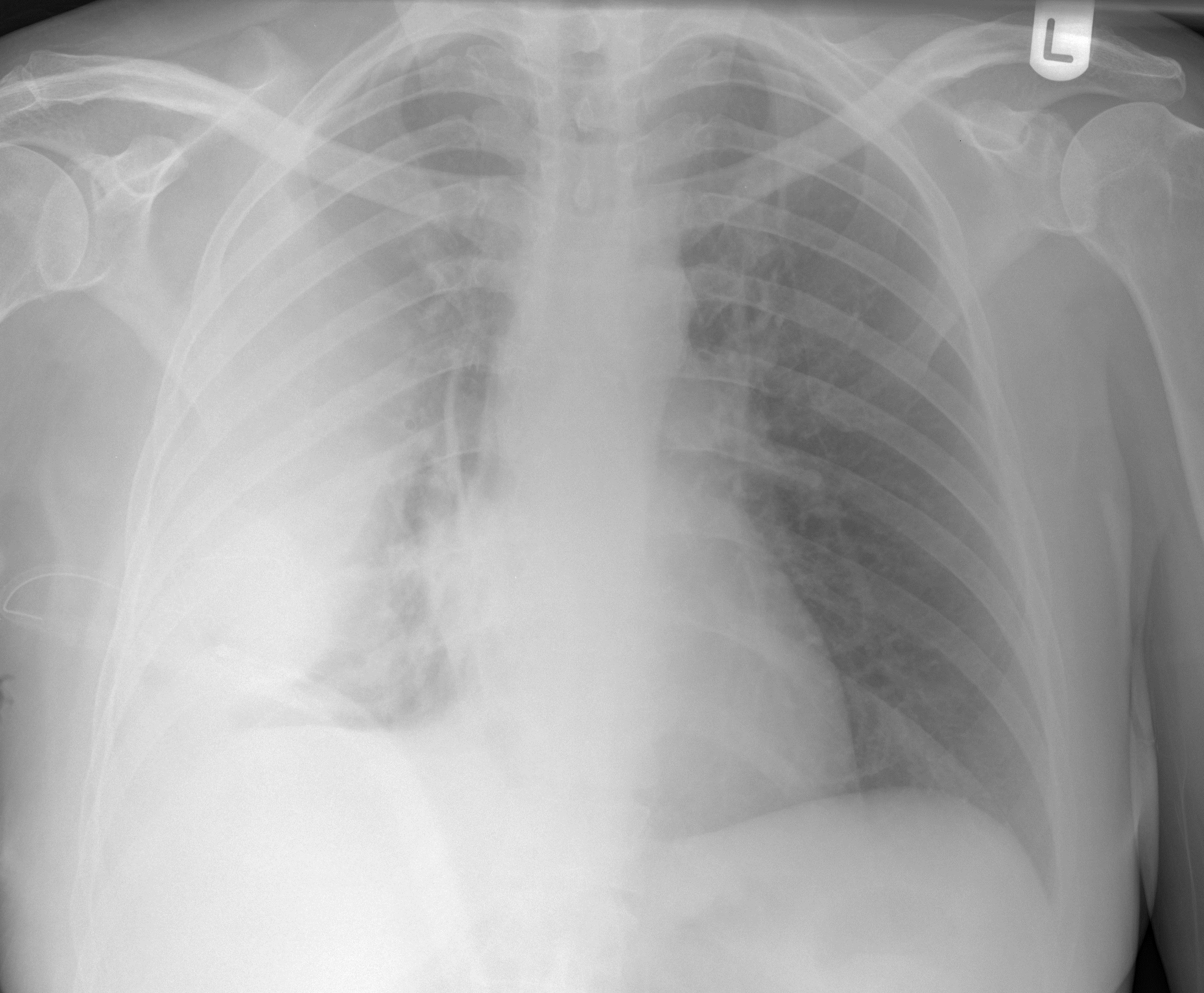
Pleural Space Infections/Empyema - The Clinical Advisor from media.clinicaladvisor.com Pleural effusion develops when more fluid enters the pleural space than is removed. What procedures and tests diagnose pleural effusions? Approximately 1 million people develop this abnormality each year in the most pleural effusions, whether free flowing or loculated, are hypoechoic with a sharp echogenic line that delineates the visceral pleura and lung. Us scan they can be identified clearly and it is very complicated.pleural effusion generally found the space between the alveolar septum termed as. Obliteration of left costophrenic angle with a wide pleural based dome shaped opacity projecting into the lung noted tracking along the cp angle and. It allows distinction between free and loculated fluid showing its extent and localization. In addition to fluid, pleural thickening, septations and calcifications can add to the functional deterioration of lungs. Effusion loculated pleural effusion pleural effusion pleural effusion (fluid around lung) secondary pleural effusion clinical information a disorder characterized by an increase in amounts of fluid within the pleural cavity.
Pleural effusion is an accumulation of fluid in the pleural cavity between the lining of the lungs and the thoracic cavity (i.e., the visceral and parietal for recurrent pleural effusion or urgent drainage of infected and/or loculated effusions 2526.
Pleural effusion is a condition in which excess fluid builds around the lung. The patient's history and physical exam may indicate a presumptive. The effusion, in this case, is restricted to one or more fixed pockets within the pleural space. In addition to fluid, pleural thickening, septations and calcifications can add to the functional deterioration of lungs. Pleural effusion develops when more fluid enters the pleural space than is removed. Pleural effusion is a common clinical problem that frequently causes dyspnoea and poor ventilatory function. Although pleural effusion volumes can be estimated by visual inspection with good correlation, some overestimation is consistently seen. This position is called lateral decubitus position. Pleura is a mesothelial lined sac that envelopes the lungs and comprises of 2 membranous walls i.e. Effusion loculated pleural effusion pleural effusion pleural effusion (fluid around lung) secondary pleural effusion clinical information a disorder characterized by an increase in amounts of fluid within the pleural cavity. Us scan they can be identified clearly and it is very complicated.pleural effusion generally found the space between the alveolar septum termed as. If you miss a tension pneumothorax you risk your patient's. The annual incidence of pleural effusion in the developed world has been estimated at 320 per 100,000 population per year 1.
Excluding the loculated effusions, the coefficient of correlation was 0969 for the right side and 0949 for the left side (p<001) loculated pleural effusion. The left lower zone is uniformly white.


0 Komentar